Research
Application of nonlinear optical microscopy for collagenous tissue study
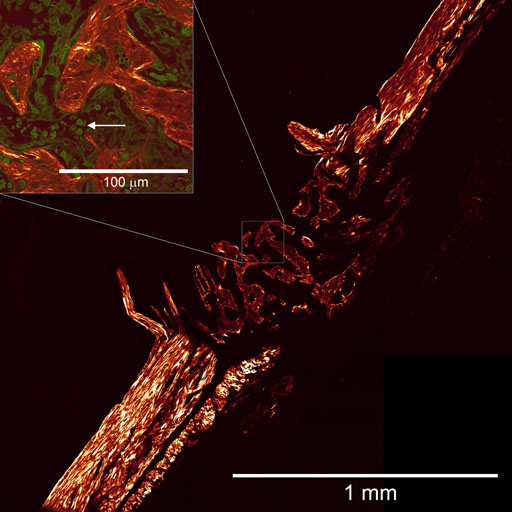
Nonlinear optical microscopy (NLOM) is an advanced optical imaging technique that produces clear images of biological specimens at the subcellular resolution level [1]. The key subsystem of NLOM is an ultrashort-pulse laser whose radiation is tightly focussed in a biological specimen. At focus, the enormous optical intensity (10^17-fold that of the sunlight for a short time interval of 100 femtoseconds) elicits nonlinear optical response resulting in photon emission at a frequency twice that of the incident photons. This process is called second harmonic generation (SHG). Simultaneous absorption of two (or several) photons followed by fluorescence emission is called multiphoton excited fluorescence (MPEF). Under typical conditions, the infrared ultrashort-pulse laser excitation is filtered, so that the SHG and MPEF signals in visible spectral range result in high-contrast en face imaging, if the focal spot is scanned across the specimen. This NLOM image formation entails the most valuable property of MPM, "optical sectioning", i.e. clearing a micron-thin image slice from the obscuring turbidity of the rest of the specimen.
We apply NLOM to image collagenous tissue, such as cartilage, bone, skin, etc., relying on the strong second-harmonic signal from collagen. In collaboration with medical and laser physics researchers from Lomonosov Moscow State University, we investigated the collagen state following the infrared laser treatment – the procedure used for cartilage reshaping [2].
NLOM imaging aided by image analysis showed great diagnostic potential, as demonstrated on the example of scar tissue classification.
1. Zipfel WR, Williams RM, Webb WW: Nonlinear magic: multiphoton microscopy in the biosciences. Nature Biotechnology 2003, 21:1368-1376.
2. Ignatieva NY, Guller AE, Zakharkina OL, Sandnes B, Shekhter AB, Kamensky VA, Zvyagin AV: Laser-induced modification of the patellar ligament tissue: comparative study of structural and optical changes. Lasers in Medical Science 2011, 26:401-413.
 |
|
Figure. NLOM (SHG) image of the unstained mouse bone fracture, showing the bone regrowth (centre). Inset: An overlay image of SHG (red) and MPF (green) zoomed area (white square). Red blood cells (arrow) are clearly observable. Preliminary results of our collaborative work with the ANZAC Research Institute investigators. |