Project 2. Nanotoxicology of sunscreens based on zinc oxide
ZnO nanomaterial affords a range of important applications due to its unique optical properties. In particular, the high optical absorption of ZnO in the most harmful UV spectral ranges of 400 nm - 315 nm (UVA) and 315 nm - 280 nm (UVB) is widely utilised in cosmetics for UV screening to protect against the radiation damage of skin. The use of ZnO nanoparticles (NP) in modern sunscreen formulations allows uniform coverage of skin surface, and renders sunscreen transparent improving overall aesthetic appeal. The cosmetic industry push toward further improvements in sunscreen transparency leads to reduction of ZnO particles mean-size to below 20 nm, although this is tempered by the safety concerns associated with ZnO NP penetration in skin. Application of ZnO-based sunscreen is generally regarded safe, if the nanomaterial stays on the skin surface, or is confined in stratum corneum (SC), the uppermost layer of skin. Despite SC is only about 10-40-micrometre thick, it provides superb protection against environmental assaul. The next underlying skin layer, termed viable epidermis, contains viable cells, which are susceptible to toxicological hazards associated with extraneous nanomaterials. It is consented that it is best to avoid penetration of ZnO NPs into viable epidermis and deeper skin layers. Hence, assessment of ZnO NP concentration in skin, including SC, viable epidermis and deeper dermal layer of skin becomes an important research focus in cosmetics, dermatology, and nanotoxicology.
A number of techniques have been demonstrated for assaying of skin ZnO nanomaterial absorption in skin, although none of these have been ideal. The emerging technique of nonlinear optical microscopy (NLOM) has demonstrated a considerable promise, as an in vivo imaging modality capable of providing clear images of the subsurface skin layers (at a depth of ≤ 200 microns in turbid biological tissue) at the sub-cellular resolution (an commercial system DermaInspect, Jen-Lab GmbH Schillerstraße 107745 Jena Germany was instrumental for these experiments). ZnO NP appeared to exhibit high NLOM-imaging contrast on the intense autofluorescence background of live skin [1].
In collaboration with the medical researchers from The University of Queensland, University of Adelaide (head, Prof Michael Roberts), Swiss researchers , University of Bern (head, Prof Martin Frenz), we carried out quantitative evaluation of the ZnO-nano distribution in skin [2].
We were trying to switch from descriptions like “bright luminescence signal” and “high concentration of ZnO nanoparticles in skin” to quantitative statements like “800 particles per m3“ that we detected at the cluster localization (demarcated by arrows in Figure). This is important for modelling of nanoparticle transdermal transport, especially in live skin, where the nanoparticle distribution in stratum corneum is the key input parameter for the model. The reported quantitative measurements will also allow determining the method threshold sensitivity, so that by stating “ZnO NPs exhibited small absorption in stratum corneum, with no detectable absorption in viable epidermis” one can infer that the amount of ZnO nanomaterial in skin is negligible to present any hazards. One of the key impact was to reassure consumers that nano ZnO unlikely to penetrate intact human skin.
Based on the existing body of evidence, it is reasonably well established that nanoparticles, including ZnO nanoparticles, of the size range greater than 15 nm, cannot pass the intact top skin layer, under conditions close to leisure and occupational practices. It is now important to establish whether this holds if sunscreen (or any other nanoparticle-based cosmetic/pharmaceutical formulations) is worn under broader range physiological conditions, for example, sweating, or damaged skin. There are a few indications that the skin permeability may alter. In vivo assessment of ZnO (and other NPs) absorption in skin becomes an indispensible tool for medical researchers to draw meaningful well-investigated conclusions.
The second key focus of our prospective research is determination of the nanoparticle permeability threshold that depends on size, charge and surface functional groups. This will create a knowledge base, from which one can design nanoparticle vehicles for transdermal drug delivery (which is proclaimed as one of the most promising drug delivery routes), or design protection means against environmental assault at the nanoscale.
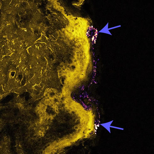
Figure. An overlay of the confocal/multiphoton image of the excised human skin. Yellow color, skin autofluorescence excited by 405 nm; purple color, ZnO NP distribution in skin (stratum corneum) excited by 770 nm, with collagen-induced faint SHG signals in the dermal layer. Arrows point to the ZnO NP high-density clusters.
Our research has attracted considerable public interest. For example, our results were communicated in various popular journals and online sources, including Science Daily, Biophotonics magazine, etc.
2. Song Z, Kelf TA, Sanchez WH, Roberts MS, Ricka J, Frenz M, Zvyagin AV: Characterization of optical properties of ZnO nanoparticles for quantitative imaging of transdermal transport. Biomedical Optics Express 2011, 2:3321-3333.